
VIP member

Thorlabs confocal microscope
Thorlabs confocal microscopy system Thorlabs' confocal laser scanning (CLS) microscopy system consists of a series of integrated imaging modules, prov
Product details
 |
|
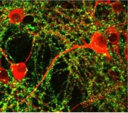

Thorlabs confocal microscope system
Thorlabs' confocal laser scanning (CLS) microscopy system consists of a series of integrated imaging modules, providing all the necessary confocal imaging tools for research laboratories. By eliminating interference signals generated outside the focal plane, confocal microscopy technology can obtain high-resolution optical slice images in thick samples and reduce background fluorescence in thin culture media. The CLS system can be integrated into almost any upright or inverted microscope, and is connected to the middle image plane (i.e. camera interface) through C-Mount threads. Maximize its application convenience, integrate it easily into existing imaging devices, and provide high-quality images. The accompanying software drives the CLS hardware through an intuitive graphical user interface, allowing for quick data recording and viewing.
ThorImageLS: A software suite guided by intuitive workflows
ThorImageLS? Developed together with multiphoton and confocal microscopy platforms to ensure seamless, logical, and intuitive integration between software and hardware. The workflow oriented interface only displays the parameters you need for each scanning series (such as Z-series for volume scanning, time series for dynamic imaging, or bleaching sequence for photoactivation/release experiments). Each software mode provides a smooth learning curve, guiding researchers step by step to complete data collection, allowing you to capture images with just a few clicks.
ThorImageLS is the complete solution for our microscope platform, which not only controls the microscope but also many accessories. The inline panel can control the position of XY and Z electric platforms, the laser power on the sample, and even the tunable coherent Chameleon? Titanium: The wavelength of sapphire lasers. High automation allows you to focus on research as much as possible without distractions.
Experimental skills
- When maximizing the depth signal-to-noise ratio, change the power according to the sample depth to minimize damage
- Customize high-speed Z-stacking or image stream acquisition parameters
- Select the area of interest for photoactivation/bleaching through an easy-to-use interface
device control
- Control the power of independent excitation lasers
- Insert/remove dichroic mirrors according to different scanning types
- Integrate active or slave TTL signals with point physiology kit
- Tune coherent Chameleon? Titanium: wavelength of sapphire laser
data analysis
- Assign a color to each detection channel
- Calculator determines the size and resolution of the image in a timely manner
- Generate three-dimensional Z-stack restoration
Product Features
- Upgrade your research grade microscope to a confocal imaging system
- Compatible with upright or inverted microscopes
- Compact and modular design
- Video rate image capture (512 x 512 pixels, speed of 30 frames per second)
- resolution
- 2048 x 2048 pixels (bidirectional)
- 4096 x 4096 pixels (unidirectional)
Product parameters
Specifications |
|
Excitation |
|
Laser Source |
1 to 4 Channels (See Table Below for Pre-Configured Options) |
Primary Dichroic Mirror |
Quad-Band Dichroic Beamsplitter |
(Other Dichroics Available upon Request) |
|
Scanning |
|
Scan Head |
Galvo-Resonant Scan Head with 8 kHz Resonant Scanner (X) and Galvo Scan Mirror (Y) |
Galvo-Resonant Scanning Speed |
30 Frames per Second at 512 x 512 Pixels |
400 Frames per Second at 512 x 32 Pixels |
|
2 Frames per Second at 4096 x 4096 Pixels |
|
Scan Zoom |
Up to 2048 x 2048 Bi-Directional Acquisition; Up to 4096 x 4096 Uni-Directional Acquisition |
1X - 16X (Continuous) |
|
Digitization / Sampling Density |
Up to 2048 x 2048 Bi-Directional Acquisition; Up to 4096 x 4096 Uni-Directional Acquisition |
Difficulty Limited Listen to Field of View (FOV) |
FN25 = 442 μm x 442 μm FOV @ 40X; FN23 = 407 μm x 407 μm FOV @ 40X |
Emission |
|
Photomultiplier Tubes (PMTs) |
Standard Multialkali or High-Sensitivity GaAsP |
Detection Channels |
1 to 4 PMTs |
Filters |
Emission Filter Set and Longpass Dichroic to Complement Multi-Channel Laser Source |
(See Table Below for? Pre-Configured Options) |
|
Laser Source Options |
||||||
Laser Source #a |
Excitation Wavelengths |
Included Emission Filters |
||||
UV |
Blue |
Green/ |
Red |
Emission Filters |
Longpass Dichroic Cutoff Wavelength(s) |
|
Orange |
(Center Wavelength/Bandwidth) |
|||||
CMLS-A |
- |
488 nm |
- |
642 nm |
525 nm/45 nm and 635 nm/Longpass |
562 nm |
CMLS-Bb |
405 nm |
488 nm |
- |
642 nm |
447 nm/60 nm, 512/25 nm, and 635 nm/Longpass |
495 nm and 538 nm |
CMLS-C |
- |
488 nm |
532 nm |
642 nm |
513 nm/17 nm, 582 nm/75 nm, and 635 nm Longpass |
538 nm and 649 nm |
CMLS-D |
- |
488 nm |
561 nm |
642 nm |
525 nm/45 nm, 607 nm/36 nm, and 535 nm/Longpass |
562 nm and 649 nm |
CMLS-Eb |
405 nm |
488 nm |
532 nm |
642 nm |
447 nm/60 nm, 513 nm/17 nm, 582 nm/75 nm, and 635 nm/Longpass |
495 nm, 538 nm, and 649 nm |
CMLS-Fb |
405 nm |
488 nm |
561 nm |
642 nm |
447 nm/60 nm, 525 nm/45 nm, 607 nm/36 nm, and 635 nm/Longpass |
495 nm, 562 nm, and 469 nm |
CMLS-Gb |
405 nm |
488 nm |
588 nm |
642 nm |
447 nm/60 nm, 525 nm/45 nm, 615 nm/24 nm, and 635 nm/Longpass |
495 nm, 605 nm, and 649 nm |
CMLS-H |
- |
488 nm |
- |
660 nm |
525 nm/39 nm and 697 nm/58 nm |
562 nm |
CMLS-I |
- |
488 nm |
- |
- |
525 nm/45 nm |
N/A |
CMLS-J |
- |
488 nm |
532 nm |
- |
512 nm/25 nm and 582 nm/75 nm |
538 nm |
>>Recommended products similar to Olympus BX51 fluorescence microscope:
 |
| Stereofluorescence microscope Model: MZX80
|
 |
| fluorescence microscope Model: MF41
|
 |
| fluorescence microscope Model: MF30
|
 |
| 5-megapixel microscope camera Model: MD50
|
Online inquiry
-
Contacts
-
Company
-
Telephone
-
Email
-
WeChat
-
Verification Code
-
Message Content
-